Computational Anatomy enables analysis of biological variability on a population. Using statistical inspired mathematical techniques, models can be built to represent the typical shape of an anatomical structure and the predominant patterns of variability in shape across a given population. These techniques allow to have a compact representation of the range of shapes in the population, which can be used to generate new virtual shapes and to predict, from sparse information, the shape of unknown ones.
Some basic research questions include:
How to create patient-specific models from sparse information, in order to minimise radiation doses to patients and optimise image segmentation procedures through patient-specific atlases?
How to model and effectively utilize population-specific models to improve patient treatments, minimise surgery Invasiveness, optimise orthopaedic implant designs?
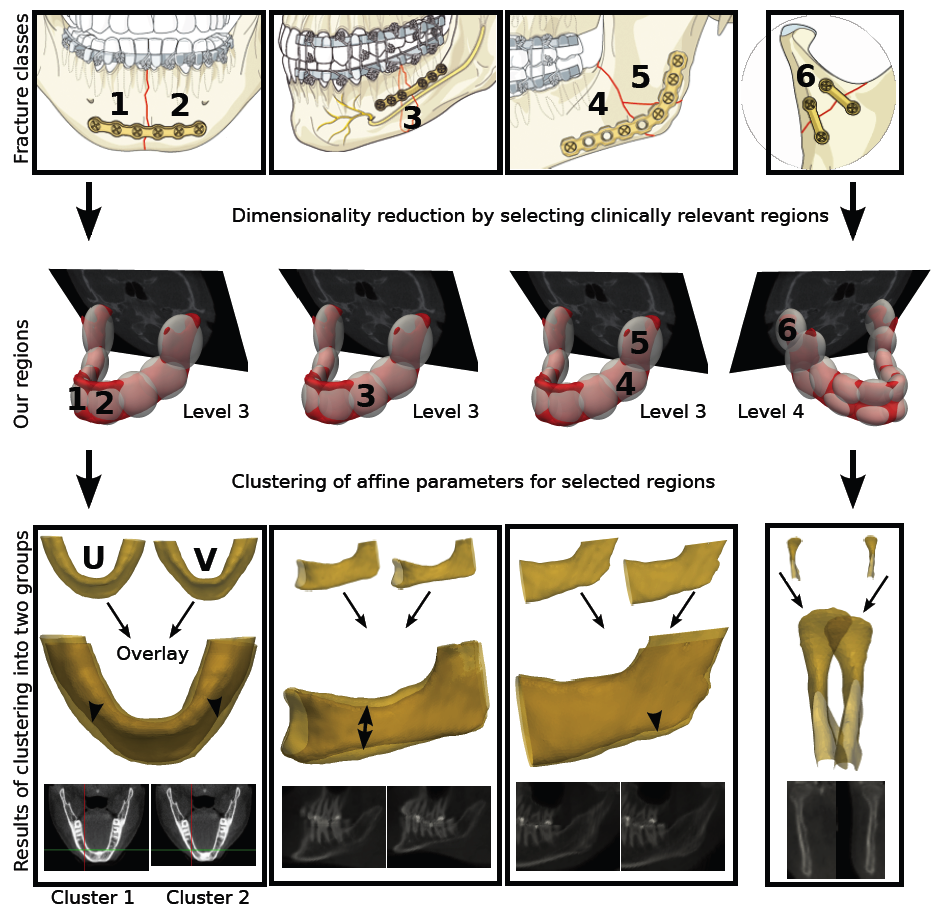
Figure: Anatomy-aware bone shape modellling. Computational anatomy based techniques are being used to model the shape variability within a population while considering clinically-relevant shape descriptors. In this example, the anatomy of the mandible is described following the AO fracture classification. The technique can be then used, for instance, for improved design of implants
The main objective of this project is to develop methodologies to perform computer-assisted orthopaedic implant design based on analysis of a given population. The outcome of this project is of great benefit to the orthopaedic implant manufacturing industry. Through the use of statistical shape analysis techniques, it is intended to guide the design of implant towards eivdence-based implant design.
The following is a video featuring one of the tools developed to automate the task of evaluating a given implant design to bone shapes.
1.
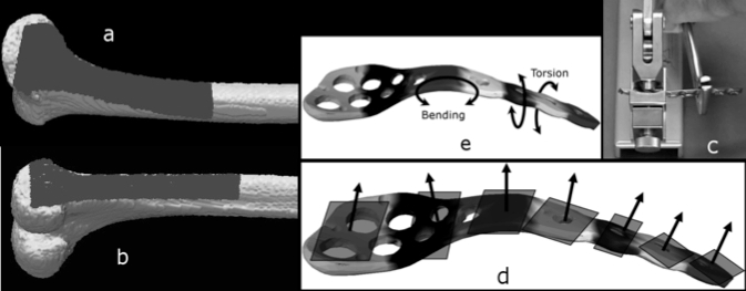
We have also developed a design framework that aims to minimize the bending and torsion deformations typically needed to adapt a given plate to a patient. The technique permits then to consider optimal fitting (minimization of distance between plate and bone surface), and optimal preoperative situation (minimization of pre-operative bending/torsion efforts). Ultimately this technology aims to guide surgeons pre-operatively to yield an optimal plate adapted to the patient's bone shape
Brain and brain tumor Imaging
PhD students: Huanxiang Lu, Stefan Bauer
Magnetic Resonance Imaging (MRI) is a powerful image modality that encompasses rich anatomical and physiological information at a high resolution. These projects aim at developing fast methods to analyze MRI images with focus on image registration and segmentation, interpretation and pattern recognition. For this, multiprocessor graphics card technologies are used, and novel methodologies are being developed to effectively assess the quality of motion correction techniques in MRI imaging. Special emphasize has given to the problem of non-rigid registration of multimodal brain images. In this sense, a novel multimodal non-rigid registration method was developed, featuring fast and accurate non-rigid registration.
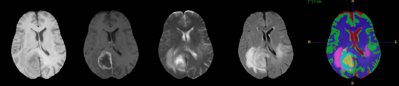
Research efforts have been put to employ these methodologies for the task of brain image segmentation in the presence of tumours. We have developed a brain segmentation methodology that permits to classify healthy and brain tumors considering the clinical imaging protocol. In addition, a finer segmentation technique based on atlas-based segmentation has been developed to deal with tumor-bearing brain images. The methodology utilizes a tumor-growth model to effectively model physiopathological-induced tissue deformations.
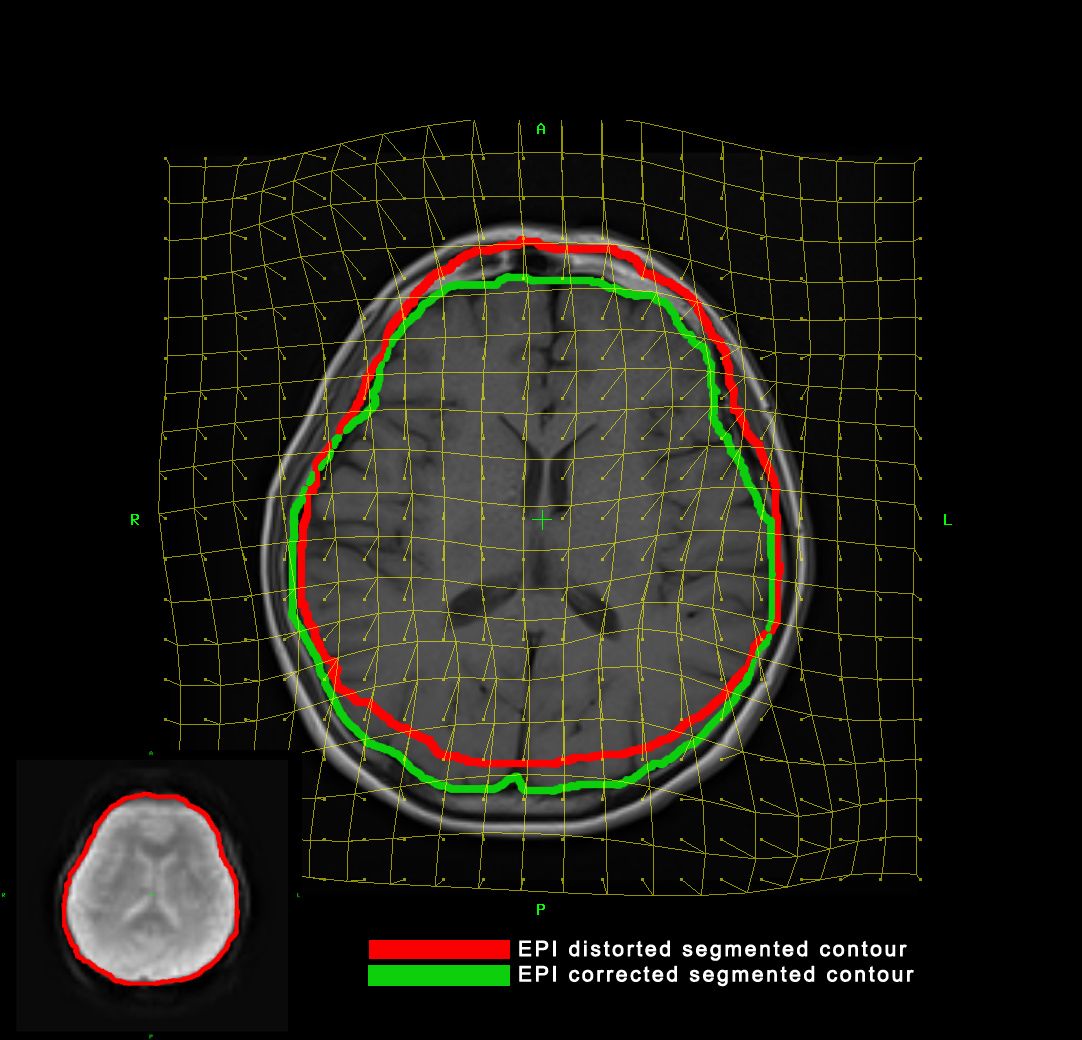
Correction of distortion in echo-planar (EPI) images (useful modality for functional neuroimaging. See bottom left corner) through combination of a synthetic deformation model and a novel multimodal diffeomorphic non-rigid image registration method. A T1 image (in the center) is used as a reference image to correct for the distortions in the EPI image. The red and green contours show the segmented EPI image before and after correction, respectively. A deformed grid has been overlaid to describe the local deformations applied to the EPI image.
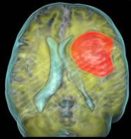
Example of brain tumor segmentation. A glioma case is segmented using an atlas-based approach and a tumor-growth simulation (work in collaboration with the computational biomechanics at the ISTB, Univ. Bern).
Multichannel tumor-bearing brain image segmentation. The clinical protocol imaging is used entirely to segment healthy (white and gray matter, and cerebrospinal fluids) and pathological tissues (necrotic, edema and active region).
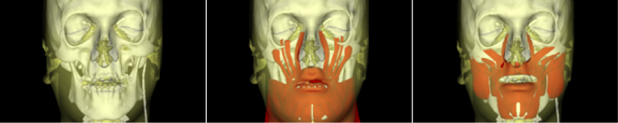
In cranio-maxilo facial surgery it is of great interest to provide surgeons with tools to predict the surgery outcome on a patient-specific basis. Simulating facial soft tissue deformations is a challenging task, moreover considering the practical requirements of computational times and easy-to-use aspects.
In this project we aim to develop fast and reliable computational methods to simulate soft tissue deformations, giving particular attention to the pre-operative and surgical pipeline.
Soft tissue simulations for Computer-Assisted Cranio Maxillo Facial (CMF) Surgery. Example case of the system used to predict soft tissue deformations after CMF surgery. Middle and Right-most image show different muscle templates used to compute the deformations. Right-most muscle model developed by Giuseppe Giovanni and Prof. Edoardo Mazza, ETHZ.
Thesis Project: Respiratory Motion Compensation in Emission
Tomography
(2003-2005)
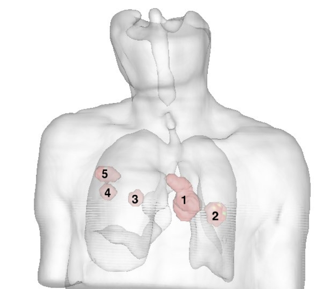
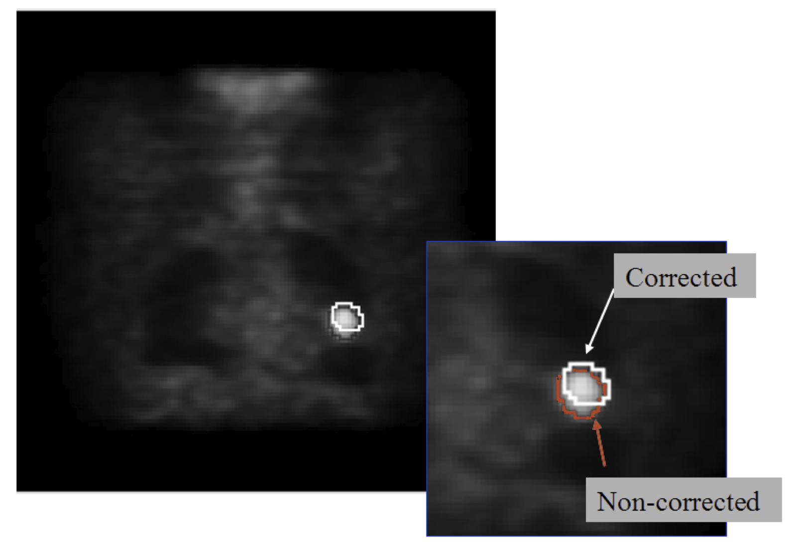
In this thesis, a new motion correction method for emission tomography was proposed. For retrospective studies and cases where no patient-specific breathing information is available, respiratory motion models were developed and used within the image reconstruction procedure in order to compensate for breathing motion, hence improving the quality of the reconstructed image.
The following animation shows the voxel model used to account for elastic deformations occured due to breathing patterns.
Two respiratory models were developed, based on a single anatomical plus physiological model, as well as a stastitical model of respiratory motion, constructed from several 4D CT scan datasets. For both cases, breathing patterns were modeled as displacement vector fields obtained through elastic registration of different states of breathing. Both models (single ans statistical) are adapted to the patient anatomy through affine and non-rigid registration, and the obtained transformations are then further used to adapt the breathing patterns, in order to create a patient-specific breathing model.
 Example of brain tumor segmentation. A glioma case is segmented using an atlas-based approach and a tumor-growth simulation (work in collaboration with the computational biomechanics at the ISTB, Univ. Bern).